In medical and clinical laboratories, the use of Amino Acid Liquid Chromatography results in highly precise determination of therapeutic compounds, metabolites, and biochemical markers. It facilitates creation of detailed patient sample profiles for research and diagnostics. The laboratory personnel prefer Amino Acid Liquid Chromatography for confirming method reproducibility, validating analytical procedures, and keeping track of sample integrity. The ultrahigh sensitivity and versatility of the apparatus permit the laboratories to cater to varied applications, thus helping hospitals and research centers to provide reliable and accurate analytical results in various fields of science.

In Amino Acid Liquid Chromatography used to analyze metabolic profiles and biomarkers during clinical research laboratories. It enables the identification of disease markers and monitoring of biochemical changes over time through the separation of small molecules and proteins. Amino Acid Liquid Chromatography also facilitates the study of drug absorption and distribution, toxicity testing, and hospital-based clinical trials and thus making it possible to monitor patient responses to therapies in great detail while at the same time ensuring the accuracy and reliability of the analytical results.

Amino Acid Liquid Chromatography is expected to have an increasing role in personalized medicine, analyzing complicated biomarkers swiftly. In the future, their application in hospitals will be centered on integrating pharmacokinetics, metabolomics, and monitoring, helping medical practitioners have access to swift and comprehensive data. The workflow in laboratories is expected to be organized.
Proper handling and care of Amino Acid Liquid Chromatography ensure continuous accuracy in the medical laboratory workflows. Cleaning of flow paths, checking detector response, and verifying pump performance are the essential maintenance tasks. Along with the column storage, solvent selection, and routine calibration, laboratory personnel must adhere to the manufacturer guidelines. Proper care enhances reproducibility, reduces downtime, and supports the consistent performance of the laboratory in hospitals and clinical research facilities.
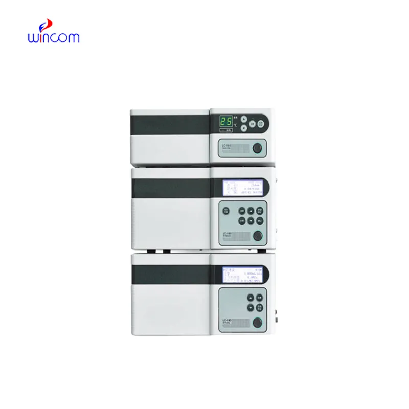
Amino Acid Liquid Chromatography is commonly employed in laboratories to separate, identify, and quantify chemical compounds. The sample mixture is put through the columns along with the stationary phases and the different components interact with the stationary phase, thus the separation is done accurately. This process not only gives high resolution but also reproducibility thus it is a must-have tool for the research works in the area of drugs, pollution, and food control. Subsequently, when coupled with sensitive detectors, Amino Acid Liquid Chromatography facilitates the precise measurement of minor concentrations. The method versatility produces so much that it has become a necessity in a routine analysis and complex research applications where it is positioned as an essential instrument in contemporary analytical chemistry and experimental workflows.
Q: What is HPLC used for in laboratories? A: HPLC turns out to be one of the most significant and essential analytical methods in laboratories equipped with the chemical compound analysis, separation, identification, and quantification of their presence in complex samples which are the research, clinical, and pharmaceutical applications. Q: How does HPLC separate compounds? A: The HPLC separation technique is based on the different affinities of the compounds to the stationary phase and mobile phase within the chromatography column. Q: Can HPLC analyze biological samples? A: Yes, it is certainly possible to carry out analyses on various biological fluids such as blood, serum, urine, etc. for the detection of metabolites, drugs, and biomarkers. Q: How often should HPLC columns be replaced? A: The replacement of the columns must be done according to the manufacturer instructions or when the performance begins to decline, which is quite usual after heavy use or contamination. Q: What detectors can be used with HPLC? A: The analysis type determines the use of, among others, UV, fluorescence, refractive index, and mass spectrometry detectors as the common detectors.
This x-ray machine is reliable and easy to operate. Our technicians appreciate how quickly it processes scans, saving valuable time during busy patient hours.
We’ve been using this mri machine for several months, and the image clarity is excellent. It’s reliable and easy for our team to operate.
To protect the privacy of our buyers, only public service email domains like Gmail, Yahoo, and MSN will be displayed. Additionally, only a limited portion of the inquiry content will be shown.
I’m looking to purchase several microscopes for a research lab. Please let me know the price list ...
I’d like to inquire about your x-ray machine models. Could you provide the technical datasheet, wa...
E-mail: [email protected]
Tel: +86-731-84176622
+86-731-84136655
Address: Rm.1507,Xinsancheng Plaza. No.58, Renmin Road(E),Changsha,Hunan,China

 af
af
 es
es
 ar
ar
 tr
tr
 sw
sw
 pt
pt
 th
th
 ur
ur
 bn
bn
 ne
ne
 vi
vi
 km
km
 lo
lo
 de
de
 ru
ru
 fi
fi
 nl
nl
 fa
fa
 fr
fr
 ko
ko