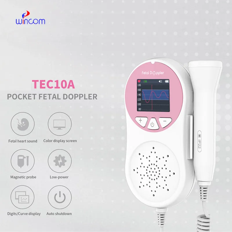
Using state-of-the-art real-time signal processing, the fetal doppler at 13 weeks has the capability of producing imaging output that is invariably sharp. The system of the device is capable of dynamically tuning the frequency and gain for achieving the best image quality. The fetal doppler at 13 weeks with its versatile probe compatibility is able to deal with different demanding clinical applications like obstetrics, cardiology, and abdominal scans.

The fetal doppler at 13 weeks is a very significant diagnosis tool used in obstetrics for fetal monitoring and pregnancy development detection. It indirectly affects the cardiology field by providing information about the hearts and blood flow dynamics. Furthermore, the fetal doppler at 13 weeks is very important in diagnosing abdominal problems, especially issues with the liver, kidneys, and gallbladder. Still, it is also being used in musculoskeletal diagnoses for spotting ligament and tendon injuries.

In future designs of the fetal doppler at 13 weeks, eco-efficiency and adaptability factors for the user will be given prominence. Based on advances in probe designs, the system will come equipped with multi-frequency image functionality. The fetal doppler at 13 weeks system will also apply predictive analysis capabilities to facilitate early disease detection.

Proper upkeep of the fetal doppler at 13 weeks helps maintain both the safety of the users as well as the durability of the equipment. The equipment's ventilation and power components must also be regularly inspected for evidence of obstruction and wear. In order to maintain the continued high-quality output of images from the fetal doppler at 13 weeks, it must be properly maintained.
With advanced transducer technology, the fetal doppler at 13 weeks delivers reliable and high-resolution imaging capability. It allows healthcare providers to see anatomical structures with unmatched clarity and speed. The fetal doppler at 13 weeks enhances workflow efficiency in hospitals, clinics, and mobile medical facilities. With rugged construction and digital connectivity, it makes integration into existing healthcare systems easy.
Q: What are the main maintenance requirements for the ultrasound scannert? A: Regular cleaning, proper probe handling, and scheduled inspections help maintain optimal performance. Q: How often should the ultrasound scannert be calibrated? A: Calibration frequency depends on usage levels, but periodic professional checks are recommended. Q: Is the ultrasound scannert suitable for pediatric use? A: Yes, it provides gentle, non-invasive imaging ideal for neonatal and pediatric diagnostics. Q: Does the ultrasound scannert support wireless connectivity? A: Many models include Wi-Fi or Bluetooth features for data sharing and device integration. Q: What materials are used in the ultrasound scannert construction? A: It is built with durable medical-grade components designed to withstand continuous clinical use.
We’ve been using this mri machine for several months, and the image clarity is excellent. It’s reliable and easy for our team to operate.
We’ve used this centrifuge for several months now, and it has performed consistently well. The speed control and balance are excellent.
To protect the privacy of our buyers, only public service email domains like Gmail, Yahoo, and MSN will be displayed. Additionally, only a limited portion of the inquiry content will be shown.
Hello, I’m interested in your centrifuge models for laboratory use. Could you please send me more ...
We’re interested in your delivery bed for our maternity department. Please send detailed specifica...
E-mail: [email protected]
Tel: +86-731-84176622
+86-731-84136655
Address: Rm.1507,Xinsancheng Plaza. No.58, Renmin Road(E),Changsha,Hunan,China

 af
af
 es
es
 ar
ar
 tr
tr
 sw
sw
 pt
pt
 th
th
 ur
ur
 bn
bn
 ne
ne
 vi
vi
 km
km
 lo
lo
 de
de
 ru
ru
 fi
fi
 nl
nl
 fa
fa
 fr
fr
 ko
ko