The fetal doppler at 7 weeks is a device that has very-sensitivity transducers, which are responsible for improving the depth of penetration and the clarity of the image. Moreover, the digital display shows anatomical structures in such a way that the human eye cannot find any fault in the accuracy of the depicted image. Besides, the device is designed for fast data storage and quick retrieval so that the healthcare provid

The fetal doppler at 7 weeks has a very wide use in radiology where it supports the guidance of non-invasive procedures. It is very important in gynecology where it is allowed to conduct reproductive system evaluations. In orthopedics, the fetal doppler at 7 weeks helps visualize muscles, joints, and tendons ensuring correct diagnostic interpretation. It is this very nature of versatility that makes it a must-have for imaging during procedures in real time.

In future designs of the fetal doppler at 7 weeks, eco-efficiency and adaptability factors for the user will be given prominence. Based on advances in probe designs, the system will come equipped with multi-frequency image functionality. The fetal doppler at 7 weeks system will also apply predictive analysis capabilities to facilitate early disease detection.

Proper care and management of the fetal doppler at 7 weeks needs to be carried out to ensure that it functions well at all times. Cleaning of the probes using a recommended disinfectant helps prevent contamination of the probe and image distortion. Storage of the fetal doppler at 7 weeks in a clean and dry place away from high temperatures helps prolong the life of the equipment.
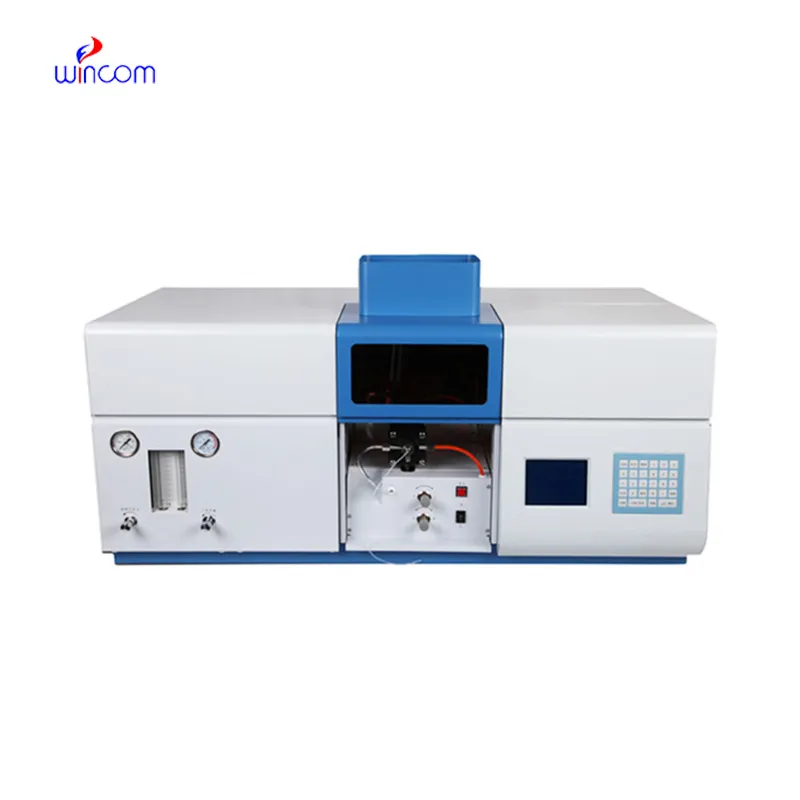
Used in hospitals and clinics, the fetal doppler at 7 weeks provides immediate visual feedback for a variety of medical evaluation uses. Converting sound waves into live images, the fetal doppler at 7 weeks allows physicians to easily detect abnormalities. The fetal doppler at 7 weeks assists with making diagnostic processes safer in addition to improving patient outcomes. It possesses an ergonomic shape alongside digital integration capabilities that support simple data sharing and medical record documentation.
Q: What imaging modes are available on the ultrasound scannert? A: It supports multiple modes such as B-mode, M-mode, and color Doppler for diverse diagnostic applications. Q: How does the ultrasound scannert improve diagnostic accuracy? A: By providing high-resolution images and real-time feedback, it enables more precise medical evaluations. Q: Can the ultrasound scannert be used in field or remote settings? A: Yes, its portable versions are designed for mobility and can be used in clinics, hospitals, or mobile healthcare units. Q: What kind of display does the ultrasound scannert use? A: It typically features a high-definition digital display that enhances image visualization and readability. Q: How is data from the ultrasound scannert managed? A: The device allows secure storage, easy access, and export of imaging data through USB or network connections.
We’ve used this centrifuge for several months now, and it has performed consistently well. The speed control and balance are excellent.
We’ve been using this mri machine for several months, and the image clarity is excellent. It’s reliable and easy for our team to operate.
To protect the privacy of our buyers, only public service email domains like Gmail, Yahoo, and MSN will be displayed. Additionally, only a limited portion of the inquiry content will be shown.
Could you please provide more information about your microscope range? I’d like to know the magnif...
I’m looking to purchase several microscopes for a research lab. Please let me know the price list ...
E-mail: [email protected]
Tel: +86-731-84176622
+86-731-84136655
Address: Rm.1507,Xinsancheng Plaza. No.58, Renmin Road(E),Changsha,Hunan,China

 af
af
 es
es
 ar
ar
 tr
tr
 sw
sw
 pt
pt
 th
th
 ur
ur
 bn
bn
 ne
ne
 vi
vi
 km
km
 lo
lo
 de
de
 ru
ru
 fi
fi
 nl
nl
 fa
fa
 fr
fr
 ko
ko