hplc laboratory is a critical technique to obtain analytical information in studies of medicines, clinical samples, and biochemistry. It isolates compounds according to their chemical characteristics, generating reproducible analytical results. Laboratory scientists use hplc laboratory to perform drug stability tests, monitor patient biomarkers, and find impurities. Its very high accuracy and flexibility allow thorough sample analysis in research, hospital, and clinical laboratory environments, thus becoming a fundamental device for assuring precision in both experimental and diagnostic results.

hplc laboratory are utilized by clinical laboratories for hormone and endocrine-related biomarker detection. It delivers trustworthy information for the diagnosis of endocrine diseases by correctly separating substances like cortisol, thyroid hormones, or insulin. Techs in a laboratory rely on hplc laboratory to give accurate and repeatable results, thereby helping doctors in individual treatment plan.

The future of hplc laboratory stresses the integration of hospital information systems and electronic medical records. The analysis of patient samples will be automatically included in the clinical workflows. Increased automation, AI-based interpretation, and better sensitivity will put hplc laboratory at the center of the laboratory operations and patient care that is focused on the patient's needs.
Preventive maintenance is hplc laboratory that play a very important role in clinical and hospital laboratories. The routine performance of flushing columns, cleaning injector valves, and monitoring pressure stability extends the life of the system. The laboratory staff is required to keep records of maintenance activities, replace consumables in a timely manner, and use solvents that are compatible. All of these practices are essential for the instruments' performance retention, lifespan extension, and high-quality analytical results, both in patient sample testing and research.
Therapeutic drug monitoring relies heavily on hplc laboratory in hospital settings. It determines the concentration of drugs in the body to guarantee efficiency and security. The laboratory staff uses it for the examination of blood, serum, or urine samples, and signifies small molecular compounds with high accuracy. By yielding consistent outcomes, hplc laboratory services the medics in changing the amounts and preventing side effects. Its use goes to hormone level testing, metabolite analysis, and pharmacokinetics research. With quick processing and accurate information, hplc laboratory is a part of the hospital patient care, making evidence-based treatment decisions possible and enhancing clinical outcomes in different departments.
Q: What types of HPLC columns are available? A: Reversed-phase, normal-phase, ion-exchange, and size-exclusion columns are the main types of columns used according to the nature of the analytes. Q: Can multiple samples be analyzed simultaneously? A: Yes, in high-throughput systems, automated sample injection and sequential analysis are among the techniques to achieve this. Q: How does temperature affect HPLC performance? A: Temperature changes can cause variations in separation efficiency and retention times; however, the majority of labs make use of precise temperature control. Q: Can HPLC be integrated with data software? A: Sure, it can be linked with laboratory software for data collection, processing, and reporting. Q: What types of laboratories use HPLC? A: HPLC is employed by hospitals, pharmaceuticals, biochemistry research, and environmental testing labs.
The water bath performs consistently and maintains a stable temperature even during long experiments. It’s reliable and easy to operate.
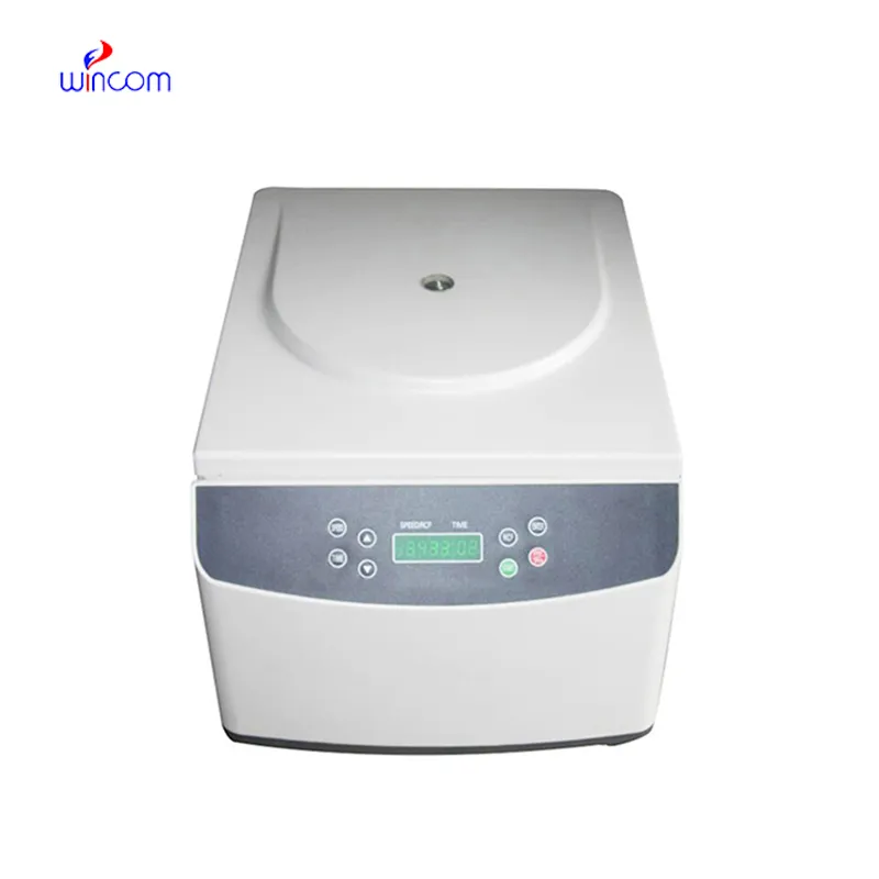
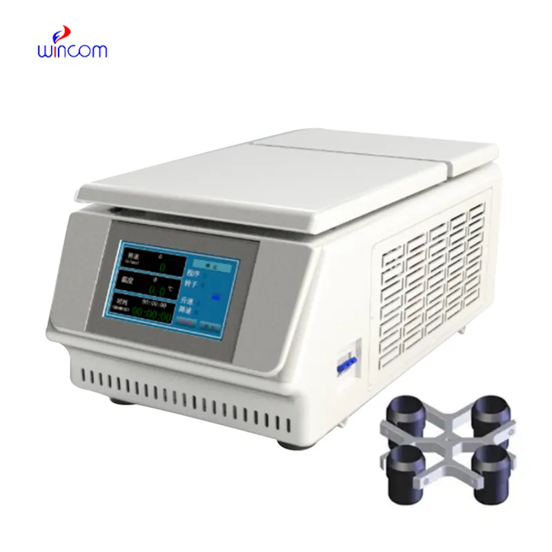
We’ve used this centrifuge for several months now, and it has performed consistently well. The speed control and balance are excellent.
To protect the privacy of our buyers, only public service email domains like Gmail, Yahoo, and MSN will be displayed. Additionally, only a limited portion of the inquiry content will be shown.
Hello, I’m interested in your centrifuge models for laboratory use. Could you please send me more ...
We’re looking for a reliable centrifuge for clinical testing. Can you share the technical specific...
E-mail: [email protected]
Tel: +86-731-84176622
+86-731-84136655
Address: Rm.1507,Xinsancheng Plaza. No.58, Renmin Road(E),Changsha,Hunan,China

 af
af
 es
es
 ar
ar
 tr
tr
 sw
sw
 pt
pt
 th
th
 ur
ur
 bn
bn
 ne
ne
 vi
vi
 km
km
 lo
lo
 de
de
 ru
ru
 fi
fi
 nl
nl
 fa
fa
 fr
fr
 ko
ko