hplc uplc enables labs to separate and analyze intricate mixtures with utmost precision. Through a seamless connection with current detectors, the method provides detailed profiling of both chemical and biological substances. The researchers and therapists trust hplc uplc for the purposes of monitoring outcomes of experiments, method development, and cross-analyses accuracy. Its strength in dealing with various kinds of samples renders it an indispensable device in both the research and the clinical settings, thus improving reproducibility and backing up the struggling with more complex scientific and medical inquiries.

The hospital laboratory technicians employ hplc uplc to quantify the quantity of proteins or peptides. This assists in the research of biomarkers, immunotherapy studies, and analysis of responses induced by novel therapies among patients. Its accuracy and sensitivity enable the obtaining of correct results, hence aiding superior research.

In hospitals and clinical research, hplc uplc techniques will get higher resolution columns and ultrafast chromatography methods more and more. It will be possible to do these innovations in a shorter time and with a more accurate result. Future hplc uplc applications will be used to identify biomarkers quickly, monitor therapies in real-time, and manage patients more efficiently in both the laboratory and clinical settings.
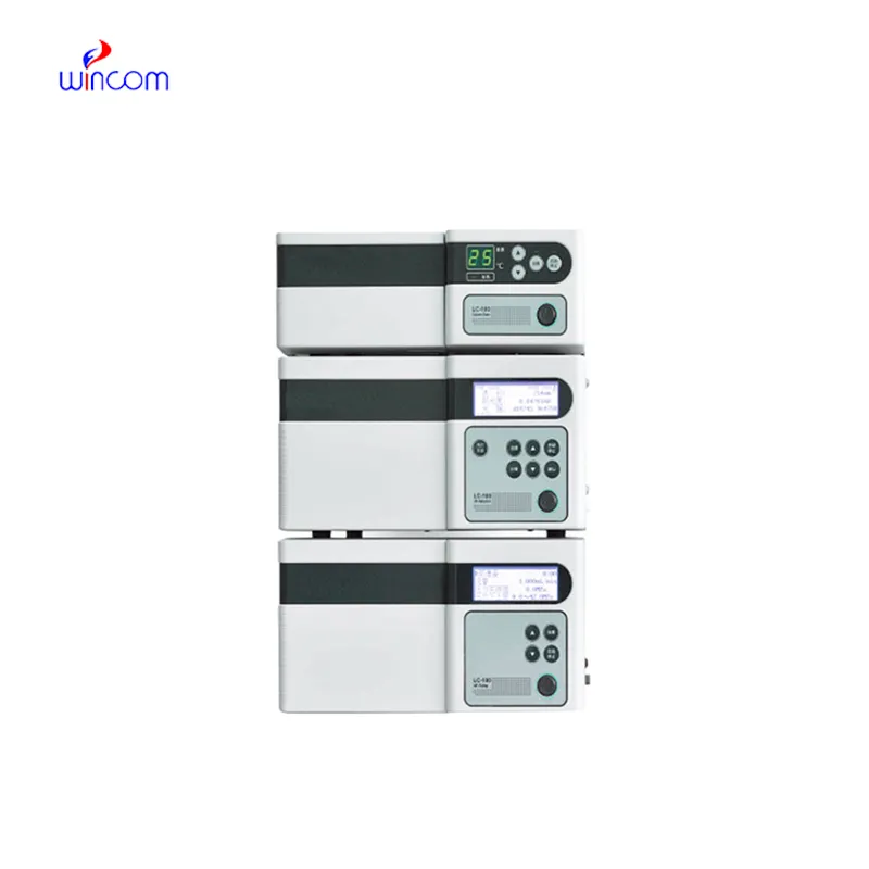
The effectiveness of a laboratory is determined by the proper maintenance of hplc uplc. If the pump seals are regularly cleaned, the flow rates are monitored, and the usage of incompatible solvents is avoided then damage to the laboratory equipment can be prevented. It is essential for the technicians to carefully examine the columns, detectors, and tubing and in case of any sign of wear to conduct the scheduled calibration. Keeping hplc uplc in their best condition guarantees reproducibility, lowers the risk of equipment breakdown, and provides continuous performance for both hospital tests and experiments.
hplc uplc is a standard method in diagnostic laboratories of hospitals to keep an eye on patients’ biochemical and therapeutic figures. It quantifies drugs, hormones, and small molecules accurately. hplc uplc speeds up the clinical decision-making processes of physicians and facilitates treatment modifications by supplying them with quick and precise results. It is used by hospital labs for basic patient testing, pharmacokinetic studies, and special analyses. The method’s high reproducibility makes certain that the outcomes are consistent, whereas its versatility allows for the support of many clinical applications. hplc uplc has turned into an irreplaceable instrument in hospital diagnostics, which not only enhances patient management but also provides healthcare professionals with thorough molecular information.
Q: What are the main parts of a microscope? A: The key components include the eyepiece, objective lenses, stage, focusing knobs, and illumination system, all working together to magnify and clarify specimens. Q: How do you clean the lenses of a microscope? A: Lenses should be cleaned using soft lens paper or microfiber cloth with a small amount of lens cleaner to avoid scratching or damaging optical coatings. Q: What magnification levels can a microscope achieve? A: Depending on the model, a microscope can typically achieve magnifications ranging from 40x to over 1000x for detailed observation of microscopic structures. Q: Why is light adjustment important in a microscope? A: Proper light adjustment ensures accurate contrast and brightness, allowing clear observation without distortion or glare during viewing. Q: Can a microscope be used for educational purposes? A: Yes, microscopes are widely used in classrooms and laboratories to teach students about biology, materials science, and microscopic analysis.
The microscope delivers incredibly sharp images and precise focusing. It’s perfect for both professional lab work and educational use.
We’ve been using this mri machine for several months, and the image clarity is excellent. It’s reliable and easy for our team to operate.
To protect the privacy of our buyers, only public service email domains like Gmail, Yahoo, and MSN will be displayed. Additionally, only a limited portion of the inquiry content will be shown.
I’d like to inquire about your x-ray machine models. Could you provide the technical datasheet, wa...
We’re interested in your delivery bed for our maternity department. Please send detailed specifica...
E-mail: [email protected]
Tel: +86-731-84176622
+86-731-84136655
Address: Rm.1507,Xinsancheng Plaza. No.58, Renmin Road(E),Changsha,Hunan,China

 af
af
 es
es
 ar
ar
 tr
tr
 sw
sw
 pt
pt
 th
th
 ur
ur
 bn
bn
 ne
ne
 vi
vi
 km
km
 lo
lo
 de
de
 ru
ru
 fi
fi
 nl
nl
 fa
fa
 fr
fr
 ko
ko