With multi-layer coated optics, the microscopic blood in urine delivers better light transmission and image contrast. Ergonomic design allows for comfortable long-term use. The smooth stage movement and fine focusing system provide sensitive slide control for accurate analysis. The microscopic blood in urine can be used with image capture systems for recording and sharing information, supporting both live observation and digital research workflows in the classroom and lab.

Versatile in use, the microscopic blood in urine has extensive usage in laboratories, universities, and manufacturing. It is used to provide precise observation of living organisms, minerals, and artificial materials. In life science research, the microscopic blood in urine helps examine cellular processes and structures of genes. Metallurgists make use of it to examine grain boundaries and fatigue cracks, while chemists make use of it to examine crystalline compounds. It is also used in the textile industry to assess fiber quality and compositional structure at high magnification.

With the progress of technology, the microscopic blood in urine will turn into a smarter and more interactive research tool. Compatibility with AI will allow it to detect patterns, recognize anomalies, and measure data automatically. The microscopic blood in urine will also make remote diagnostics possible, where the samples from every corner of the world can be diagnosed remotely by specialists. Advances in imaging sensors and optical systems will provide better depth resolution and faster capture rates. These will expand the uses of the microscopic blood in urine in medicine, nanotechnology, and education.

Cleaning, checking, and storing the microscopic blood in urine with care is part of taking care of them. Dust accumulation can impact both optical and mechanical performance, and thus covering the microscopic blood in urine when idle is inevitable. Avoid handling objective lenses with unmasked fingers to prevent oil smudges and residues. Remove immersion oil instantly after observation. The microscopic blood in urine are kept in a controlled, temperature-stable environment. Periodic focus and illumination system calibration ensures image quality in the long term.
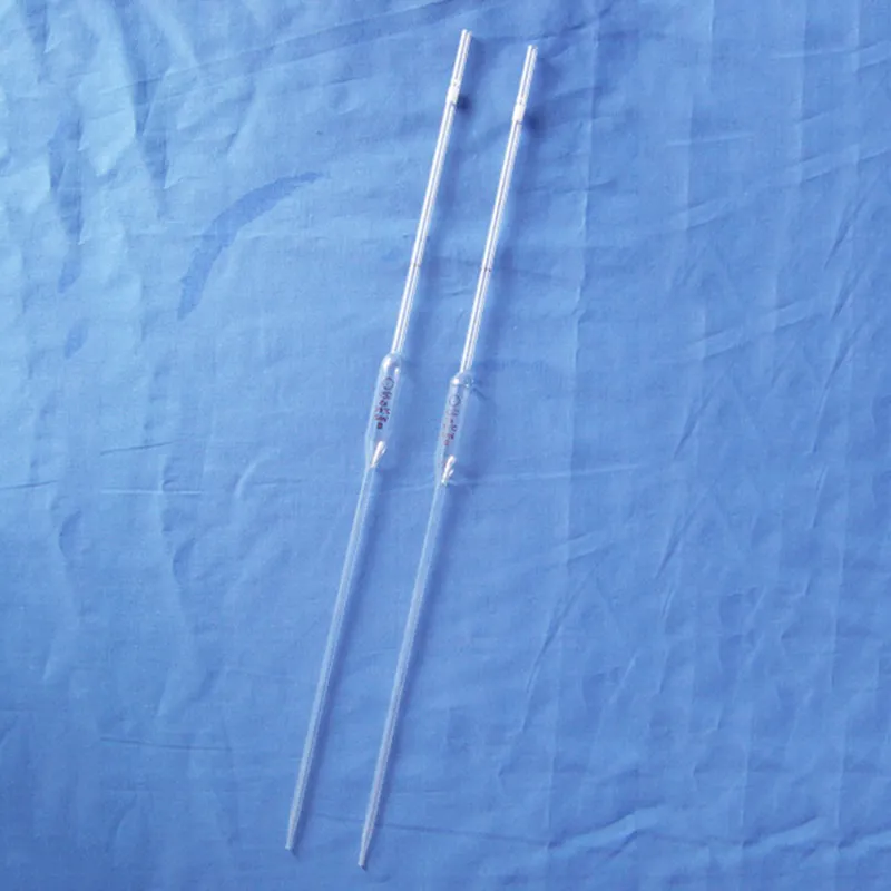
A microscopic blood in urine is able to closely study microorganisms, tissue, and materials and is thus a fundamental instrument in laboratories and classrooms. It operates by bending light or electron rays to enlarge specimens to appear gigantic many times magnification. The microscopic blood in urine has been enhanced with developments in optics to enable brighter, clearer, and digital-imaging-assisted magnification. In academic research work as well as industrial inspection, a microscopic blood in urine enables accurate analysis, recording, and examination of complex microscopic realms.
Q: What distinguishes a digital microscope from a traditional one? A: A digital microscope integrates cameras and imaging software, enabling users to view, capture, and analyze images directly on a computer or monitor. Q: How can vibration affect a microscope? A: Vibration can cause image blur or misalignment, so the microscope should always be placed on a stable, vibration-free surface. Q: What safety measures should be taken when using a microscope? A: Avoid touching optical parts with fingers, use slides carefully, and ensure electrical components are safely connected before operation. Q: Why is immersion oil used in some microscopes? A: Immersion oil increases the refractive index between the lens and specimen, improving resolution and brightness at higher magnifications. Q: How can you prevent mold growth in a microscope? A: Store the microscope in a low-humidity environment and use desiccants or dehumidifiers to keep optical components dry and mold-free.
This x-ray machine is reliable and easy to operate. Our technicians appreciate how quickly it processes scans, saving valuable time during busy patient hours.
This ultrasound scanner has truly improved our workflow. The image resolution and portability make it a great addition to our clinic.
To protect the privacy of our buyers, only public service email domains like Gmail, Yahoo, and MSN will be displayed. Additionally, only a limited portion of the inquiry content will be shown.
We’re currently sourcing an ultrasound scanner for hospital use. Please send product specification...
Hello, I’m interested in your centrifuge models for laboratory use. Could you please send me more ...
E-mail: [email protected]
Tel: +86-731-84176622
+86-731-84136655
Address: Rm.1507,Xinsancheng Plaza. No.58, Renmin Road(E),Changsha,Hunan,China

 af
af
 es
es
 ar
ar
 tr
tr
 sw
sw
 pt
pt
 th
th
 ur
ur
 bn
bn
 ne
ne
 vi
vi
 km
km
 lo
lo
 de
de
 ru
ru
 fi
fi
 nl
nl
 fa
fa
 fr
fr
 ko
ko