monolithic hplc column offers high resolution separation of complex samples in clinical, pharmaceutical, and hospital laboratories, thereby supporting advanced laboratory workflows. It allows performing an in-depth analysis of drugs, metabolites, and small biomolecules. monolithic hplc column is used by laboratory staff for research validation, patient monitoring, and method development. Its precision, speed, and adaptability make analytical efficiency greater and at the same time, make consistent and reproducible results which in turn, strengthen laboratory operations in the areas of healthcare and scientific environments.

Biochemical and clinical laboratories use monolithic hplc column to examine plasma or serum metabolites for disease research. It isolates and measures the amounts of small molecules participating in metabolism thus shedding light on patient conditions. The method is commonly employed in metabolic studies and experimental clinical trials conducted in hospitals.

The forthcoming breed of monolithic hplc column will put a spotlight on intelligent instruments that are connected with cloud-based surveillance. Through this monitoring, hospitals will be able to gain a remote view of laboratory activities and the results of sample analysis. Lab productivity will be greatly increased by the upcoming monolithic hplc column, and together with the new features, patient testing and therapy monitoring even in difficult clinical settings will be more accurate.

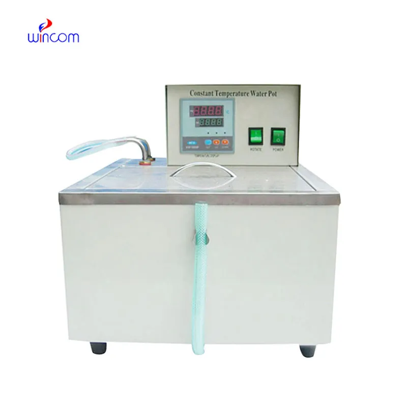
The effectiveness of a laboratory is determined by the proper maintenance of monolithic hplc column. If the pump seals are regularly cleaned, the flow rates are monitored, and the usage of incompatible solvents is avoided then damage to the laboratory equipment can be prevented. It is essential for the technicians to carefully examine the columns, detectors, and tubing and in case of any sign of wear to conduct the scheduled calibration. Keeping monolithic hplc column in their best condition guarantees reproducibility, lowers the risk of equipment breakdown, and provides continuous performance for both hospital tests and experiments.
Therapeutic drug monitoring relies heavily on monolithic hplc column in hospital settings. It determines the concentration of drugs in the body to guarantee efficiency and security. The laboratory staff uses it for the examination of blood, serum, or urine samples, and signifies small molecular compounds with high accuracy. By yielding consistent outcomes, monolithic hplc column services the medics in changing the amounts and preventing side effects. Its use goes to hormone level testing, metabolite analysis, and pharmacokinetics research. With quick processing and accurate information, monolithic hplc column is a part of the hospital patient care, making evidence-based treatment decisions possible and enhancing clinical outcomes in different departments.
Q: What is HPLC used for in laboratories? A: HPLC turns out to be one of the most significant and essential analytical methods in laboratories equipped with the chemical compound analysis, separation, identification, and quantification of their presence in complex samples which are the research, clinical, and pharmaceutical applications. Q: How does HPLC separate compounds? A: The HPLC separation technique is based on the different affinities of the compounds to the stationary phase and mobile phase within the chromatography column. Q: Can HPLC analyze biological samples? A: Yes, it is certainly possible to carry out analyses on various biological fluids such as blood, serum, urine, etc. for the detection of metabolites, drugs, and biomarkers. Q: How often should HPLC columns be replaced? A: The replacement of the columns must be done according to the manufacturer instructions or when the performance begins to decline, which is quite usual after heavy use or contamination. Q: What detectors can be used with HPLC? A: The analysis type determines the use of, among others, UV, fluorescence, refractive index, and mass spectrometry detectors as the common detectors.
The centrifuge operates quietly and efficiently. It’s compact but surprisingly powerful, making it perfect for daily lab use.
I’ve used several microscopes before, but this one stands out for its sturdy design and smooth magnification control.
To protect the privacy of our buyers, only public service email domains like Gmail, Yahoo, and MSN will be displayed. Additionally, only a limited portion of the inquiry content will be shown.
We’re looking for a reliable centrifuge for clinical testing. Can you share the technical specific...
I’m looking to purchase several microscopes for a research lab. Please let me know the price list ...
E-mail: [email protected]
Tel: +86-731-84176622
+86-731-84136655
Address: Rm.1507,Xinsancheng Plaza. No.58, Renmin Road(E),Changsha,Hunan,China

 af
af
 es
es
 ar
ar
 tr
tr
 sw
sw
 pt
pt
 th
th
 ur
ur
 bn
bn
 ne
ne
 vi
vi
 km
km
 lo
lo
 de
de
 ru
ru
 fi
fi
 nl
nl
 fa
fa
 fr
fr
 ko
ko