The pink fetal doppler has been designed considering the needs of modern health care, providing uninterrupted performance with its rapid image acquisition and high-definition visualization. The robust outer casing and sophisticated temperature control system will make sure that the device continues to work and be trustworthy. Also, the pink fetal doppler is a device that aids the long-term data archiving process for efficient medical record management.

In emergency departments, the pink fetal doppler is used for instant imaging to easily spot internal wounds and bleeding. It supports the doctor with the abdominal trauma and chest condition diagnosis. Moreover, the pink fetal doppler provides assistance in rural and field medical practice, delivering consistent imaging in areas with poor medical facilities.

The next-generation pink fetal doppler solutions come with better processing capabilities and intelligent algorithms that improve the clarity of images in addition to lessening reliance on operators. The aspect of augmented reality will change the world of surgical operations. The pink fetal doppler solutions will also change the face of delivering healthcare by facilitating quicker and more accurate diagnoses.

The daily upkeep of the pink fetal doppler involves cleaning, inspection, and proper storage. The removal of the gel residue from the probes should be accomplished as soon as the analysis has been carried out. The cooling vents of the device should always be unblocked. The pink fetal doppler needs annual professional servicing in order to remain accurate.
The pink fetal doppler uses state-of-the-art ultrasound technology to deliver real-time imaging for diagnostic and monitoring purposes. It aids physicians in assessing organs, blood vessels, and soft tissue with unmatched clarity. The non-surgical device is an important tool for guiding medical procedures and making precise diagnoses. The pink fetal doppler combines portability with precision, rendering it extremely useful in routine exams as well as emergency applications.
Q: What imaging modes are available on the ultrasound scannert? A: It supports multiple modes such as B-mode, M-mode, and color Doppler for diverse diagnostic applications. Q: How does the ultrasound scannert improve diagnostic accuracy? A: By providing high-resolution images and real-time feedback, it enables more precise medical evaluations. Q: Can the ultrasound scannert be used in field or remote settings? A: Yes, its portable versions are designed for mobility and can be used in clinics, hospitals, or mobile healthcare units. Q: What kind of display does the ultrasound scannert use? A: It typically features a high-definition digital display that enhances image visualization and readability. Q: How is data from the ultrasound scannert managed? A: The device allows secure storage, easy access, and export of imaging data through USB or network connections.
The delivery bed is well-designed and reliable. Our staff finds it simple to operate, and patients feel comfortable using it.
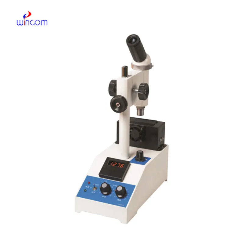
I’ve used several microscopes before, but this one stands out for its sturdy design and smooth magnification control.
To protect the privacy of our buyers, only public service email domains like Gmail, Yahoo, and MSN will be displayed. Additionally, only a limited portion of the inquiry content will be shown.
I’m looking to purchase several microscopes for a research lab. Please let me know the price list ...
Could you please provide more information about your microscope range? I’d like to know the magnif...
E-mail: [email protected]
Tel: +86-731-84176622
+86-731-84136655
Address: Rm.1507,Xinsancheng Plaza. No.58, Renmin Road(E),Changsha,Hunan,China

 af
af
 es
es
 ar
ar
 tr
tr
 sw
sw
 pt
pt
 th
th
 ur
ur
 bn
bn
 ne
ne
 vi
vi
 km
km
 lo
lo
 de
de
 ru
ru
 fi
fi
 nl
nl
 fa
fa
 fr
fr
 ko
ko