Designed with a compact and intelligent design, the the first x ray machine invented offers various modes of imaging such as full body scans and localized scans. The device supports digital viewing stations where the images can be viewed remotely by radiologists. The the first x ray machine invented assists in improved diagnostic operations by providing easy-to-handle mechanisms and sound imaging consistency.

In the hospital and clinic setting, the the first x ray machine invented is utilized for chest imaging, exposing respiratory and cardiovascular pathologies. It is widely employed to monitor pneumonia, tuberculosis, and cardiac enlargement. The the first x ray machine invented is also important in dental and maxillofacial examinations, providing precise visual markers in treatment planning.

Emerging technologies in the the first x ray machine invented will deliver hybrid imaging capabilities combining X-rays with additional modalities including ultrasound or CT overlays. This will deliver even superior diagnostic information. The the first x ray machine invented will also employ environmentally friendly components to reduce environmental footprint.

Maintenance of the the first x ray machine invented requires close attention to mechanical, electrical, and imaging parts. Regular visual examination catches wear or damage early. The the first x ray machine invented must be cleaned using non-abrasive substances, and filters or protective covers periodically replaced. Preventive maintenance minimizes downtime and provides reliable diagnostic results.
In today's healthcare system, the the first x ray machine invented continues to be an integral part of diagnostic imaging. The the first x ray machine invented provides precise visual data that helps in disease detection and assessment of an injury. The the first x ray machine invented has digital sensors and the capability to improve images. The the first x ray machine invented helps in quick and effective medical imaging.
Q: How is patient safety ensured during x-ray exams? A: Safety is maintained through minimal radiation doses, shielding equipment, and adherence to strict exposure guidelines. Q: What should be done if the x-ray image appears unclear? A: The operator should check positioning, exposure levels, and detector condition before repeating the scan under safe and controlled settings. Q: Can an x-ray machine detect metal implants or devices? A: Yes, x-ray machines can clearly show metallic objects such as implants, prosthetics, or surgical tools within the scanned area. Q: Are portable x-ray machines as effective as stationary ones? A: Portable x-ray machines are effective for bedside or emergency imaging, offering flexibility though with slightly lower image power compared to stationary units. Q: How is radiation exposure monitored for staff using x-ray machines? A: Staff wear dosimeters that record cumulative exposure levels, ensuring they remain within regulated safety limits throughout their work.
We’ve been using this mri machine for several months, and the image clarity is excellent. It’s reliable and easy for our team to operate.
The hospital bed is well-designed and very practical. Patients find it comfortable, and nurses appreciate how simple it is to operate.
To protect the privacy of our buyers, only public service email domains like Gmail, Yahoo, and MSN will be displayed. Additionally, only a limited portion of the inquiry content will be shown.
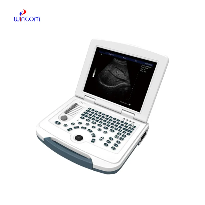
We’re currently sourcing an ultrasound scanner for hospital use. Please send product specification...
We are planning to upgrade our imaging department and would like more information on your mri machin...
E-mail: [email protected]
Tel: +86-731-84176622
+86-731-84136655
Address: Rm.1507,Xinsancheng Plaza. No.58, Renmin Road(E),Changsha,Hunan,China

 af
af
 es
es
 ar
ar
 tr
tr
 sw
sw
 pt
pt
 th
th
 ur
ur
 bn
bn
 ne
ne
 vi
vi
 km
km
 lo
lo
 de
de
 ru
ru
 fi
fi
 nl
nl
 fa
fa
 fr
fr
 ko
ko